 What is breast thermography?
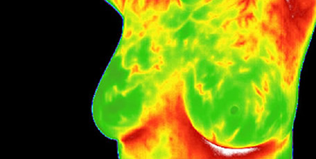
The use of a Digital Infrared Imaging is based on the metabolic activity and vascular circulation in both pre-cancerous tissue and the area surrounding a developing breast cancer is almost always higher than in normal breast tissue.
In an ever-increasing need for nutrients, cancerous tumors increase circulation to their cells by holding open existing blood, vessels, opening dormant vessels, & creating new ones. This process frequently results in an increase in regional surface temperatures of the breast.
Thermography uses ultra-sensitive medical infrared camera and sophisticated computers to detect, analyze, and produce high-resolution images of these temperature variations. Because of the extreme sensitivity, these temperature variations may be among the earliest signs of breast cancer and/or pre-cancerous state of the breast.
Just as unique as a fingerprint, each patient has a particular infrared map of their breast. Any modification of this infrared map on images taken over months or years, may constitute an early sign of an abnormality. However, if a pathology is suspected, this information is used to recommend further examination & tests.
Your history, & images are taken by an RN & then read by certified physicians of DITI and are kept on file for several years, for comparison.
Early Detection Means Life
Breast cancer is the most common cancer in women, and the risk increases with age. Risk is also higher in women whose close relatives have had the disease. Women without children, and those who have had their first child after the age of 30. Current research indicates that 1 in every 8 women in the US will get breast cancer in their lifetime.